
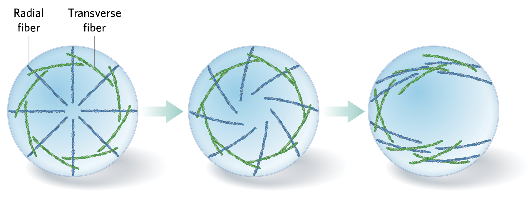
Individual cells are chiral: in addition to showing up-down and front-back asymmetries, they are also asymmetric along the left-right axis. One of the possible ways this chirality could get established inside cells is through the spontaneous self-organization of the actin filaments making up the cell cytoskeleton. In one study a few years ago, Yee Han Tee and colleagues plated fibroblasts individually onto circular adhesive islands that forced each cell to maintain a circular shape, and imaged the movements of two sets of actin fibers in each cell: radial fibers, which stretch inward from focal adhesions on the cell membrane, and transverse fibers, which are arranged perpendicularly to the radial fibers and seem to physically interact with them. In the first few hours of cytoskeleton development, these fibers formed a radially symmetrical pattern (left), but after about three hours, the radial fibers began to tilt, dragging the transverse fibers sideways and causing a swirling pattern to emerge (middle). Then, at around the 11-hour mark, the swirling broke into a linear pattern of fibers stretching across the cell (right).
Read the full story.