 2012 Labbies | Video Finalists | Image Finalists
See the finalists for best image and vote for your favorite!
Giant Unilamellar Liposome
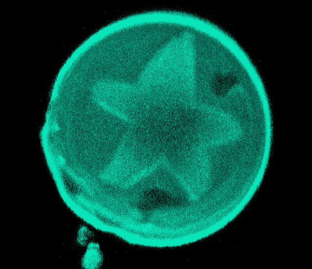
Luis Bagatolli, a biophysicist at the University of Southern Denmark, captured this image of a giant unilamellar liposome (40 µm in diameter) using laser scanning confocal fluorescence microscopy. The liposome is composed of ceramide, cholesterol, and the glycerophospholipid POPC. The star shaped region is formed by a ceramide-enriched two-dimensional crystal embedded in the lipid bilayer.
Water Bear
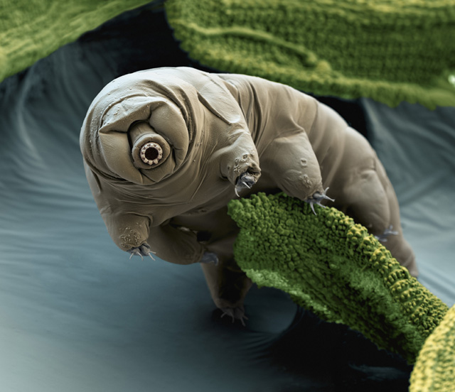
This color enhanced scanning electron micrograph (SEM) of a water bear (Macrobiotus sapiens) was captured by Eye of Science—a two-person team of photographer Oliver Meckes and biologist Nicole Ottawa. Water bears are tiny invertebrates that live in aquatic and semi-aquatic habitats such as lichens and damp moss.
Hippocampus

This high resolution image of the...
Electric Ray

This photograph of an electric ray was snapped by Hari Mohan Saxena in November 2011 during a visit to the Seattle Aquarium in Washington State. Saxena is a professor of immunology at the Guru Angad Dev Veterinary & Animal Sciences University in India, and likes to practice nature photography as a hobby.
Luna Moth

This scanning electron micrograph (SEM) of the antenna of the Luna Moth (Actias luna) was taken by scientific photographer and high school physics teacher Ted Kinsman. The antenna of A. luna, a moth native to North America, is one of the most sensitive chemical detectors known in the insect world.
2012 Labbies | Video Finalists | Image Finalists


